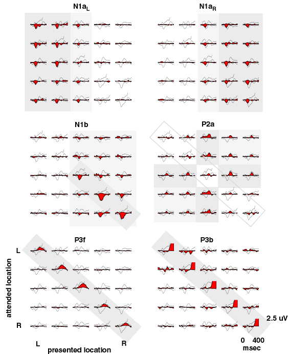
Figure 3.
Envelopes of the six largest independent components (black
outline, filled with red, from all 31 channels) (cf. Fig. 1c above),
superimposed on the mean response envelopes for all 25
Presented/Attended Location conditions. Note the systematic differences
between the sets of conditions in which the different components are
active: N1aL and N1aR (top row) are evoked by left and right visual
field stimuli, but do not appear to depend in any simple way on the
attended location. Both also respond to midline stimuli. N1b (center
left) responds to right visual field stimuli. Its amplitude is enhanced
both in attended locations and to a lesser extent in nearby right
visual field locations (locations (4,5) and (5,4)). P2a, by contrast
(middle right),
has little or no response to targets (diagonal traces).
Its amplitude is generally largest to nontarget midline stimuli.
Components P3f (lower left) and P3b
(lower right, amplitude clipped)
account for overlapping early and middle portions of the late positive
complex (Makeig et al., J. Neurosci., 1999) in responses to target stimuli
presented at attended locations.
Next