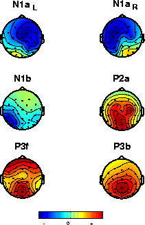
Figure 2b. Scalp maps of the largest six independent components, individually scaled (green represents zero weight). Relative locations of the electrodes are shown by small dots. Color polarities are chosen to represent the signs at their time point of maximum projection (red positive, blue negative with respect to the reference). Note the bilateral near-symmetry of the two early-N1 components (top row).
Next